animal cell under microscope 100x
See how a generalized structure of an animal cell. The cell wall is distinctly visible around each cell.

Typical Plant Cell 100x Dissection Connection Plant Cell Typical Plant Cell Cell
The sample was then stained with methylene blue so the cells could be viewed at 40x 100x and 400x.

. The images of Paulownia wood hair and frogs blood were captured with a high power compound microscope using a Nikon camera adapter. And Animal Cells SAFETY. Typical animal cell Center 100x.
Animal Cell Under Microscope. Cell had a cytoplasmcell membrane and a nucleus hard to see. Nerve cell under the microscope - Abstract blue dots on white background.
Cells have been stained to help. Be sure to label the chloroplasts the cell membrane and the cell wall. Structure Morphology and Classification.
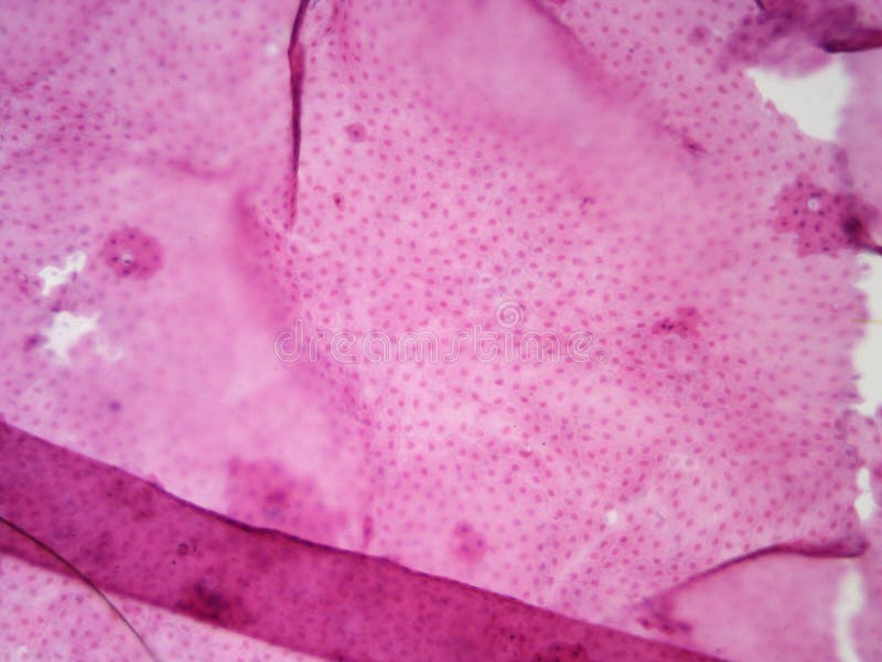
Certain parts of the cell are also clearly distinguishable with or without staining making the activity even. Focus at 100x and re center so that you are focused on the more square meristem cells. Under the microscope plant cells are seen as large rectangular interlocking blocks.
Human blood smear under 100X light microscope with abnormal red blood cells call Hypochromic and Target cells and normal white blood cell call Neutrophils Selective focus. The typical animal cell can be seen here. Cell is a tiny structure and functional unit of a living organism containing various parts known as organelles.
As such they are not plants animal or fungi. In particular they share some characteristics of both plants and animals. Animal Cell Under Microscope 10X.
In the lab we cut an onion and removed a tiny portion of the inside where cells can be viewed. Thus light microscopes allow one to visualize cells and their larger components such as nuclei nucleoli secretory. The cell wall is somewhat thick and is seen rightly when stained.
Compound Microscope 40x 100x Microscope slides Cover slips Pipette droppers Elodea Toothpicks with flat side Food Coloring Handouts with labeled plant cell. They are all typical elements of a cell. Under the microscope animal cells appear different based on the type of the cell.
What can you see in an animal cell under a light microscope. Built and used the first single lens microscope. In animal cells youll see a round shape with an outer cell membrane and no cell wall.
The following images simulate what would be seen at each magnification. Differences between animal and human hair. In this lab we took a sample of our own cells by scraping the inside of the cheek with a toothpick.
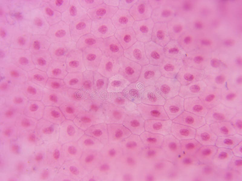
Add a drop of methylene blue solution on to the smear and gently place a cover slip on top to cover the stain and the cells any excess solution can be removed by touching one side of the slide with a paper towel or blotting paper. The compound microscope typically has three or four magnifications - 40x 100x 400x and sometimes 1000x. The granulated area is the cell Cytoplasm while the huge round part is the Nucleus.
At 40x magnification you will be able to see 5mm. Photo about microscope help stained nucleus visible magnification visibility cytoplasm membrane center cells cell compound. Huge collection amazing choice 100 million high quality affordable RF and RM images.
Up to 15 cash back Find the perfect animal cell and microscope stock photo. Take the time to go over safety guidelines. Microscope with 40X 100X and 400X magnification Microscope Slides.
Find Human Blood Smear Under 100x Light stock images in HD and millions of other royalty-free stock photos illustrations and vectors in the Shutterstock collection. The animal cell structure is the most prominent in human cheek cells. Diameter of the field of view compound microscope showing the 10x ocular eyepiece and four objectives 4x 10x 40x and 100x.
Hair can be matched by other characteristics that can be viewed under a compound microscope. Learn the structure of animal cell and plant cell under light microscope. The cytoplasm is also lightly stained containing a darkly stained nucleus at the periphery of the cellMay 26 2021.
Typical animal cell Center 40x. Our hair grows from follicles located under the skin and has two. Cell is a tiny structure and functional unit of a living organism containing various parts known as organelles.
Use the coarse adjustment knob to focus. At 100x magnification you will be able to see 2mm. The onion skin cell an example of a plant cell.
And search more of iStocks library of royalty-free stock images that features 2015 photos available for quick and easy download. There are various tasks done by a cell to complete them as the. To identify plant and animal cells you must use a microscope with at least 100x magnification power.
In both the plant and animal cells the individual chromosomes are no longer. When observing onion cells there is the Cell Surface Membrane which is present in all living cells. IStock Plant Cells Under Microscope100x Stock Photo - Download Image Now Download this Plant Cells Under Microscope100x photo now.
Hair under a compound microscope. View the leaf under low medium and high power objectives and then draw the cells in Figure 22 along with any organelles you can see. Onion epidermal cells appear as a single thin layer and look highly organized and structured in terms of shape and size.
Frozen felt tissue under the microscope 100x Human cells. Place the leaf on the slide along with a drop of water and then cover with a coverslip. No need to register buy now.
Place the slide on the microscope for observation using 4 x or 10 x objective to find the cells. In animal cells youll see a round shape with an outer cell membrane and no cell wall. Gm485125830 1200 iStock In stock.
Cell membrane nucleus and cytoplasm are all visible. While they can manufacture their own food a characteristic seen in plants they are also capable of movement. To identify plant and animal cells you must use a microscope with at least 100x magnification power.
Place the slide on the stage under low power. Please provide the students with all the tools they need to do the activities safely. 1 667 Animal Cell Microscope Photos Free Royalty Free Stock Photos From Dreamstime There are one or more.
Set up your microscope place the onion root slide on the stage and focus on low 40x power. Observing onion cells under a microscope is a fun and easy activity for students and hobbyists alike. Observe each of the prepared bacteria plant and animal under 100x magnification.
Move your slide so that your field of view is centered on the root tip. Euglena are single celled organisms that belong to the genus protist.

All The Templates You Can Download Microscopic Photography Microscopic Microscope
Typical Animal Cell Center 100x Stock Photo Image Of 100x School 152965862

Plant Stem Cross Section Microscopic Photography Dna Art Microscopic
Typical Animal Cell Center 400x Stock Image Image Of Visible Compound 152965979

Award Winning Images Of Really Tiny Things On Earth Microscopic Photography Microscopic Images Electron Microscope Images

Pin On Microscopic Inspiration For Printmaking Class
Ranunculus Root Cross Section Root Wikipedia Plant Science Science Notes Plants

Plant Cell Images Plant Cell Microscopic Photography

Various Fresh Water Diatoms 100x Diatom Single Celled Cell Wall

0002 Microscopic Photography Microscopic Biology Art

Plant Cell Under The Microscope 1 Microscopic Photography Plant Cell Microscopic Images

Kids Science Microscope Activity Yeast Microscope Activity Science For Kids Homeschool Science Experiments

Ceriodaphnia Microscopic Photography World Photography Macro Photography

Motor Neuron Cell Body Dendrites And Axon 100x Also Shows Motor Neuron Neurons Patterns In Nature

Axolot Amphibians With Liver Cells 100x Stock Photo Picture And Royalty Free Image Image 154834747

Retina Under The Microscope Microscope Human Tissue Microscopy

Celery Petiole Microscopic Cells Things Under A Microscope Rocks And Crystals

Microscope Image Of African Violet Flower Petal Cells Violet Flower Microscopic Photography Flower Petals

Onion Root Meristem Mitosis Plant Cell Microscopy